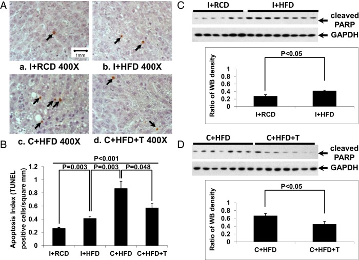
Figure 4.
Hepatocyte apoptosis. A, By terminal deoxynucleotidyl transferase-mediated deoxyuridine 5-triphosphate nick end labeling staining, more apoptotic hepatocytes (black arrows) were detected in the C+HFD group compared with other groups. B, Quantification of the apoptotic cells in liver showed that rats fed a HFD had increased hepatocyte apoptosis, which was further elevated by castration (P = .003) and reduced by T replacement (P = .048). C and D, By Western blotting, HFD led to higher levels of cleaved PARP compared with the RCD group in intact rats (Figure 4C, P < .05), and T replacement attenuated the increased PARP cleavage in hepatocytes induced by a HFD in the castrated group (panel D, P < .05). GAPDH was used as loading control.

