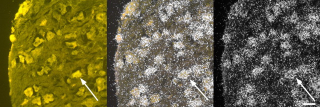
Fig. 6.
Photomicrographs of a DRG containing fluorescently labeled neurons as well as hybridization with the AR probe. The photomicrograph to the left shows cells labeled with fluorescent traces under UV illumination. The photomicrograph to the right shows the same tissue viewed under dark-field illumination to illustrate the AR probe hybridization. The photomicrograph in the center shows an overlay of the left and right photomicrographs to demonstrate that fluorescent cells express AR mRNA. Arrows point to a representative cell that shows both florescence and silver grain accumulation. Reference bar, 40 μm.

