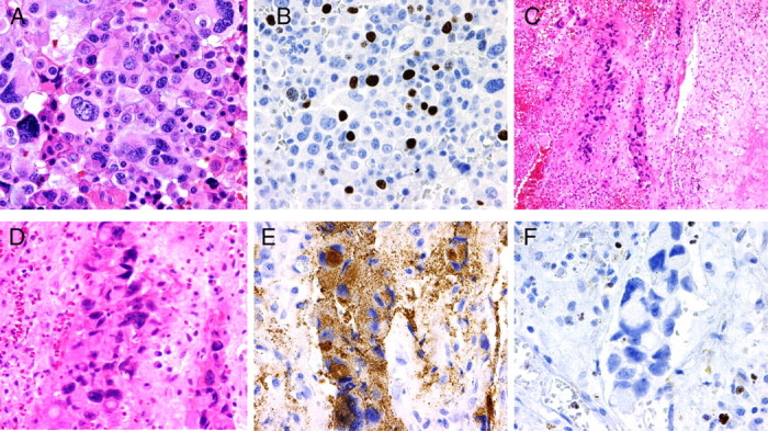
Fig. 4.

Pre- and posttemozolomide treatment specimens from case 2. A and B, Surgical specimen before GammaKnife radiation and temozolomide treatment showing extensive cellular atypia (A) and high Ki-67 labeling index (18%) (B). C–F, Surgical specimen after treatments and collected during CSF leak correction showed focal islands of tumor cells entrapped on fibrous connective tissue with intense inflammatory reaction (C). Extensive cellular pleomorphism was present (D), but adenoma cells were still immunoreactive for ACTH (E). Ki-67 labeling was practically absent on tumor cells, but present in inflammatory cells seen on the specimen (F).
