
Fig. 4.
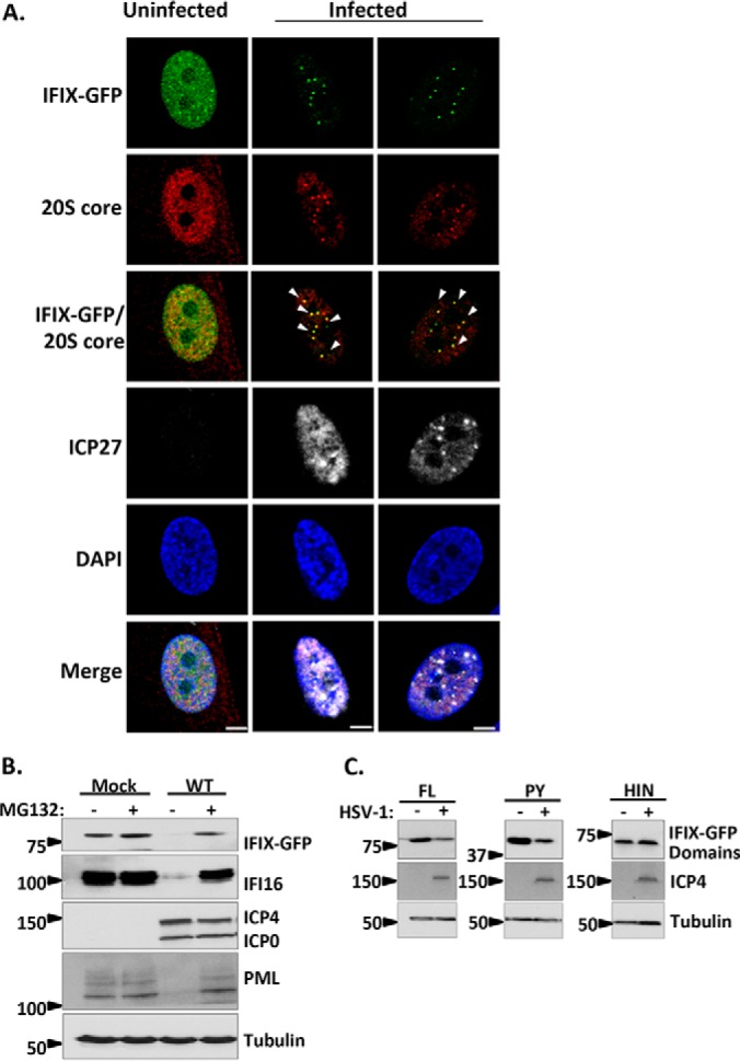
IFIX is targeted for proteasome-dependent degradation during infection with HSV-1. A, direct- and immunofluorescence microscopy during WT HSV-1 infection displaying IFIX-GFP puncta localizing to nuclear proteasomes. The 20S core subunit of the proteasome is in the red channel, and ICP27 as a marker for infection is in white. m.o.i.: 10, 3.5 hpi. Bar, 5 μm. B, Western blot of IFIX-GFP stable HFFs during WT HSV-1 infection with and without proteasome inhibitor MG132 treatment. ICP4 and ICP0 are markers for infection. IFI16 and PML are positive controls. IFIX levels decrease during infection but are rescued by MG132 treatment. m.o.i.: 10, 6 hpi. C, Western blot of 293Ts transfected with IFIX-GFP domain constructs reveals the pyrin domain of IFIX is the target for degradation. FL, full length; PY, pyrin domain; HIN, HIN200 domain. ICP4 is marker for infection. m.o.i.: 10, 6 hpi. See supplemental Fig. S1 for exposures of the intact Western blot membranes.