Fig. 6.

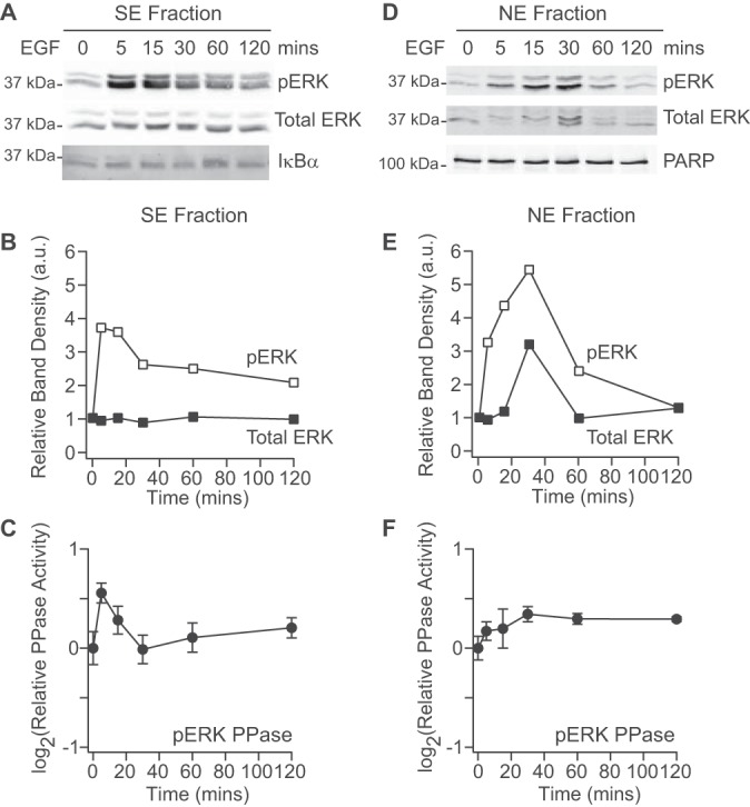
EGF stimulates subcellular ERK phosphorylation and ERK phosphatases. A–B, Immunoblots for phospho-ERK and total ERK in the SE fraction (A) were quantified by densitometry (B) with IκBα used to confirm equal loading. C, ERK2 phosphatase activity quantified in the SE fraction. D–E, Immunoblots for phospho-ERK and total ERK in the NE fraction (D) were quantified by densitometry (E) with PARP used to confirm equal loading. F, ERK2 phosphatase activity quantified in the NE fraction. Phosphatase activities are shown as the means ± standard error of n = 4 biological replicates. Cells were stimulated with 100 ng/ml of EGF for the indicated times.
