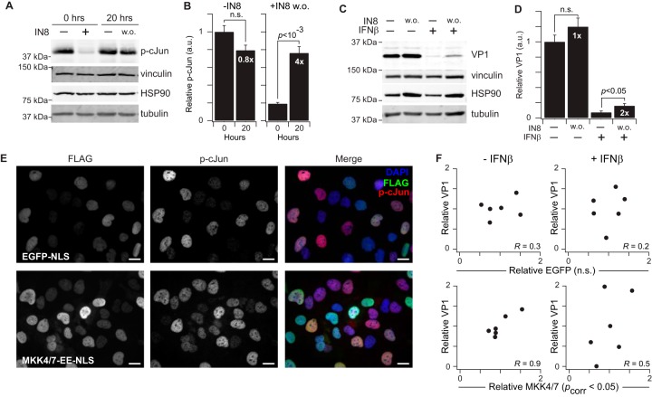
Fig. 9.
Nuclear JNK1 activity promotes CVB3 protein synthesis in infected AC16-CAR cells. A–B, Immunoblots for p-cJun before and after JNK-IN-8 (IN8) washout (w. o.) (A) were quantified by densitometry (B) with vinculin, HSP90, and tubulin used to confirm equal loading. Fold change in p-cJun relative to the corresponding pre-washout condition is shown in white. C–D, Immunoblots for VP1 with or without IN8 washout, IFNβ treatment, or both in CVB3-infected cells (C) were quantified by densitometry (D) with vinculin, HSP90, and tubulin used to confirm equal loading. Fold change in VP1 relative to the corresponding pre-washout condition is shown in white. E, Immunofluorescence of EGFP-NLS or MKK4/7-EE-NLS and p-cJun in AC16-CAR cells. Scale bar is 20 μm. F, Correlation plots between relative FLAG and VP1 abundance in infected AC16-CAR cells overexpressing EGFP-NLS (top panels) or MKK4/7-EE-NLS (bottom panels). IN8 data are shown as the means ± standard error of n = 4 biological replicates. Differences in p-cJun (B) and VP1 (D) abundance were assessed by Student's t test. FLAG-VP1 Pearson correlations (R) were assessed after Fisher Z transformation and Fisher's method of combined probabilities for each cell line.

