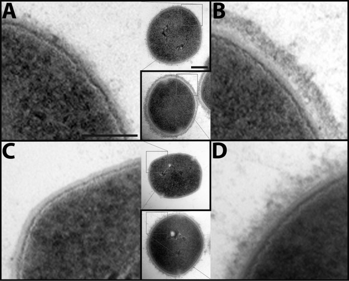
Fig. 5.
Electron micrographs of the S. pyogenes surface with and without adsorbed human plasma proteins. S. pyogenes cells from the same samples used for SRM were fixed and protein interaction networks is shown using electron microscopy: A, The wild-type strain SF370; B, SF370 with plasma adsorbed to surface; C, The SF370 M1 deletion mutant ΔM1; D, ΔM1 with plasma adsorbed to surface. The electron micrographs show a close-up of the bacterial surface, whereas the inserts depict one complete bacterium. Scale bars, 100 nm (close-ups) and 200 nm (inserts).

