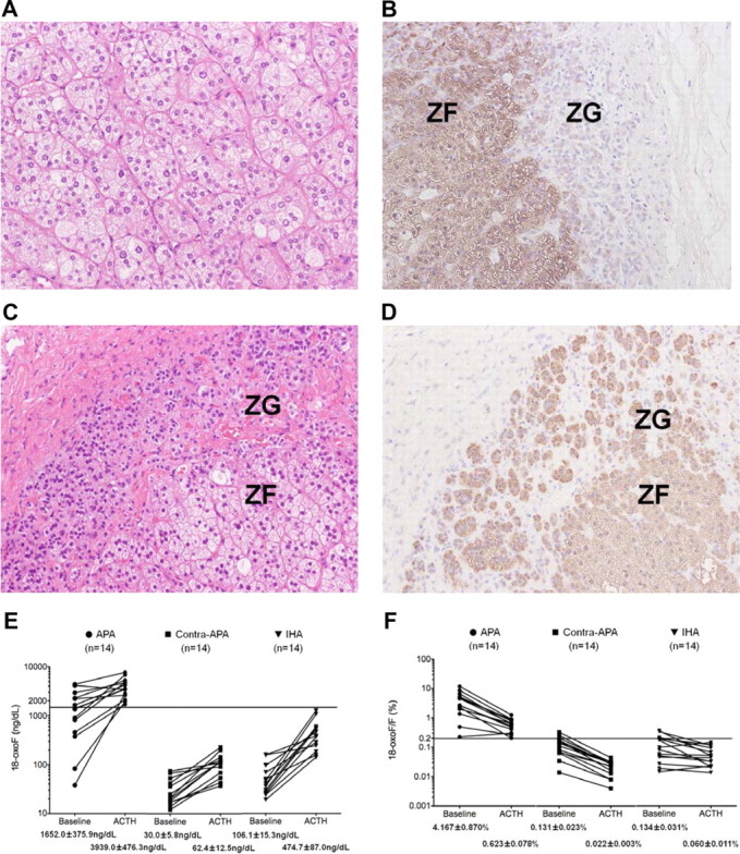
Fig. 1.

A and B, Hematoxylin-eosin staining of the APA demonstrated the presence of predominantly clear cells (A), and immunohistochemical localization of 3β-HSD (B) demonstrated low expression in adjacent hyperplastic ZG (case 6). C and D, Hematoxylin-eosin staining (C) and immunohistochemical localization of 3β-HSD (D) showing the hyperplasia and distinct immunoreactivity of 3β-HSD in the ZG of the IHA adrenal gland (case 16). E and F, The levels of 18-oxoF (E) and the ratio of 18-oxoF/F (panel F) in adrenal vein samples of APA (14 samples), the APA contralateral adrenal (14 samples), and IHA (14 samples) both before and after ACTH stimulation. Before ACTH stimulation, the mean levels of 18-oxoF and the mean ratios of 18-oxoF/F were significantly higher in blood-draining APA (1652.0 ± 375.9 ng/dl and 4.167 ± 0.870%, respectively) than in those of their contralateral adrenal glands (30.0 ± 5.8 ng/dl and 0.131 ± 0.023%, respectively) and the adrenal glands associated with IHA (62.4 ± 12.5 ng/dl and 0.134 ± 0.031%, respectively) (P ≤ 0.05). After ACTH stimulation, the mean levels of 18-oxoF and the mean ratios of 18-oxoF/F were significantly higher in blood-draining APA (3939.0 ± 476.3 ng/dl and 0.623 ± 0.078%, respectively) than in those of the contralateral-APA adrenal glands (106.1 ± 15.3 ng/dl and 0.022 ± 0.003%, respectively) and adrenal glands with IHA (474.7 ± 87.0 ng/dl and 0.060 ± 0.011%, respectively) (P ≤ 0.05). The horizontal lines show 1500 ng/dl (E) and 0.200% (F), respectively.
