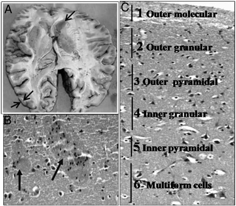
Figure 1.
A, Area showing sites of collection of gray and caudate tissue from the brain. B and C, Hematoxylin and eosin-stained photomicrographs from 2 tissues showing mature neurons embedded in a background of glial cells and neurofibrillary material with 2 patches of striatopallidal/pencil fibers (arrows) in the caudate nucleus (B) and organization of neuron into 6 layers (outer molecular, outer granular, outer pyramidal, inner granular, and multiform layer) in the gray matter (C) (magnification, ×100).

