
Figure 4. Inhibition of NTE activity represses GnRH-induced membrane recycling in LβT2 cells.
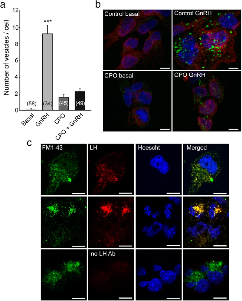
Membrane recycling was studied in LβT2 cells using the membrane dye FM1–43FX. A and B, Under basal conditions, the FM dye shows very little incorporation into the cell. When cells are stimulated with GnRH, the FM dye gets rapidly incorporated into vesicular-like structures (GnRH group in A, and control GnRH in B), indicating an increase in membrane dynamics. When NTE activity is blocked with CPO, the stimulatory effect of GnRH on vesicular recycling is inhibited. C, Vesicular structures labeled with FM1–43 contain LHβ as detected by immunofluorescence. In A, columns are means, and vertical bars are SEMs. ***, P < .001 vs all other groups (ANOVA followed by the Student-Newman-Keuls multiple-comparison test for unequal replications). Numbers in bars indicate the number of cells analyzed per treatment. B, High magnification of a representative group of cells for each treatment showing the FM1–43FX dye (labeling recently formed vesicles, in green), WGA (labeling glycoproteins of the cell membrane, in red), and Hoechst 33258 (labeling cell nuclei, in blue). Scale bars, 10 μm. C, Examples of cells with colocalization of the FM1–43FX dye (green) and LHβ immunoreactivity (red). Nuclei were identified by staining with Hoechst 33258 (blue). Scale bars, 10 μm.