
Figure 2.
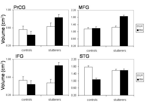
ROI analysis Mean WM volumes (and standard errors of the mean as vertical bars) in the precentral gyrus (PrCG), middle frontal gyrus (MFG), inferior frontal gyrus (IFG), and the superior temporal gyrus (STG) broken down for the left (open bars, LH) and right (filled bars, RH) hemisphere. The STG comprises Heschl's gyrus and the planum temporale. The volume measures are expressed as arbitrary values because these measures were obtained from brains transformed into the MNI space.