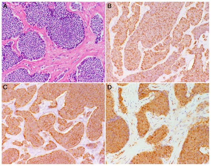
Figure 2. Tumor immunohistochemistry.
A, Hematoxylin and eosin-stained section of pancreatic NET (20 × magnification). B, Immunohistochemistry for chromogranin A (20 × magnification; brown color). C, Immunohistochemistry for synaptophysin (20 × magnification; brown color). D, Immunohistochemistry for PTEN showing strong cytoplasmic staining of tumor cells (brown color) with absent nuclear staining.

