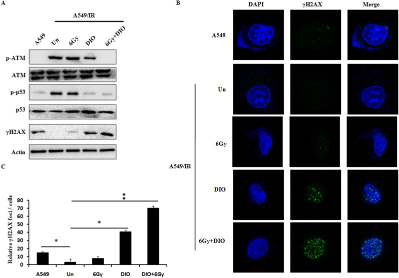
Fig 4. Effects of DIO on the DNA damage in A549/IR cells.
(A) Reduced phosphorylation of ATM and p53, and increased γH2AX expression level upon DIO treatment with or without RT. Cell lysates were processed for the indicated proteins by immunoblotting. β-actin expression shows the equal loading. (B) γH2AX foci status was investigated by using a confocal analysis for different treatment. Representative images are shown. (C) Quantitative data of γH2AX foci are summarized. Bars represent the means ± SD of triplicate samples. *P < 0.05, **P < 0.01.

