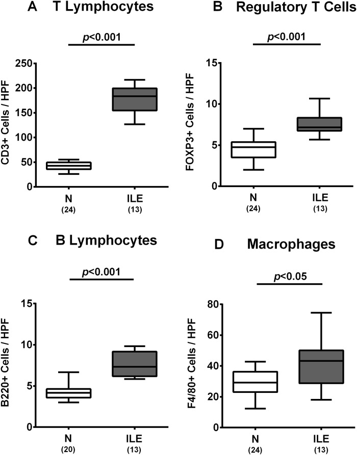
Fig 3. Small intestinal immune cell responses in human microbiota associated mice suffering from acute ileitis.
Human microbiota associated (hma) mice were perorally infected with T. gondii strain ME49 to induce acute ileitis (ILE; grey boxes). Noninfected hma mice served as controls (N, white boxes). The average numbers of ileal (A) T lymphocytes (positive for CD3), (B) regulatory T cells (positive for FOXP3), (C) B lymphocytes (positive for B220), and (D) macrophages (positive for F4/80) from six high power fields (HPF, 400 x magnification) per animal were determined microscopically in immunohistochemically stained ileal paraffin sections at day 7 post ileitis induction. Box plots represent the 75th % and 25th % percentiles of the medians (black bar inside the boxes). Total range and significance levels (p-values) determined by the Student’s t test and Mann-Whitney U test and numbers of mice (in parentheses) are indicated. Data shown were pooled from three independent experiments.

