
FIGURE 1. Polysomnographic Example of OSA.
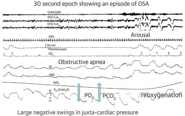
First tracing is chin electromyogram, second and third are electroencephalogram, fourth is electrocardiogram, fifth and sixth are airflow measured by thermocouple (fifth) and CO2 (sixth), seventh and eighth are rib cage (RC, seventh) and abdominal (ABD, eighth), ninth is oxyhemoglobin saturation measured by pulse oximetry, and tenth is esophageal pressure. Please note that during obstructive apnea, airflow is absent while breathing effort continues. Breathing resumes with the onset of arousal. Reprinted with permission from Javaheri (8). OSA = obstructive sleep apnea.