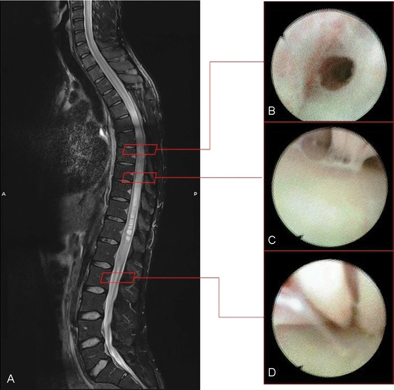
Fig. 1.

(A) Midline sagittal T2-weighted preoperative spine MRI showing syringomyelia. (B) Endoscopic imaging: the endoscope through the perimedullar fibrous septae consequent the arachnoiditis. (C) Endoscopic evidence of the fibrous adherences stretched between the cord and the arachnoid layer. (D) Endoscopic view of the roots of the cauda equina and the fibrous septae between them. MRI, magnetic resonance imaging.
