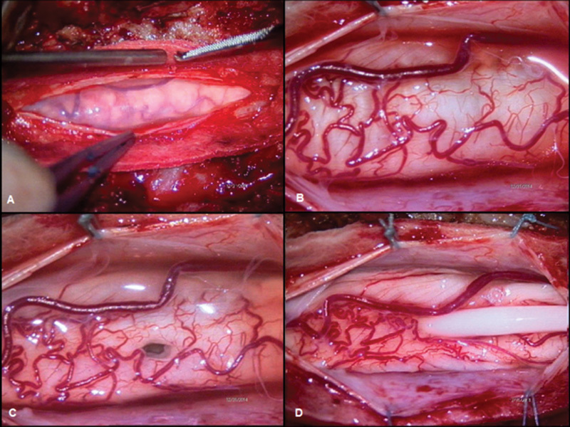
Fig. 3.

Intraoperative imaging. (A) Evidence of thickened spinal arachnoid layer. (B) Posterior view of the enlarged spinal cord. (C) A 6-mm myelostomy opened on the posterior midline. (D) The shunt in its definitive position. It is evident the decompression of the spinal cord after the drainage of the syringomyelic cavity.
