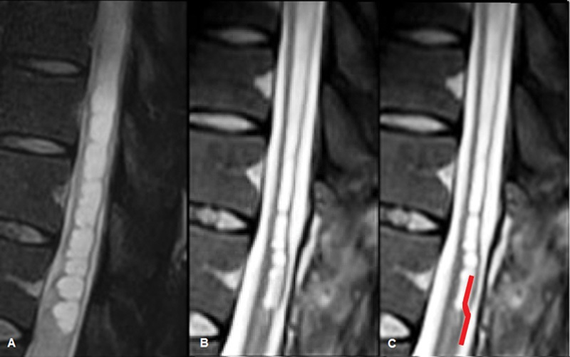
Fig. 5.

(A) Midline sagittal T2-weighted preoperative spine MR image of the syrinx. (B) Midline sagittal T2-weighted image from the spine MRI performed 40 days after the operation showing the reduction of the syrinx diameter. The device is barely evident in the lower part of the syrinx. (C) Same image as in B where the position of the silicon shunt has been artificially enhanced. MR, magnetic resonance; MRI, magnetic resonance imaging.
