Figure 2.
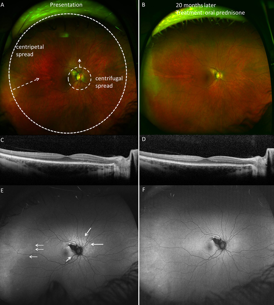
Centrifugal and centripetal spread at presentation and follow-up in subject 3. A. Fundus image of the right eye demonstrating pinpoint zone of hypopigmented outer retinal lesions surrounding optic nerve. B. Resolution of acute changes seen 20 months later, after a course of oral prednisone. C. Spectral domain optical coherence tomography (SD-OCT) image shows ellipsoid zone (EZ) and external limiting membrane (ELM) disruption in the nasal parafovea. D. SD-OCT at 20 months shows normalization of EZ and ELM disruptions outside area of retinal pigment epithlium atrophy. E. Ultra-wide-field fundus autofluorescence (UWFFAF) shows both centrifugal spread of the hyperautofluorescent lesions and centripetal spread from the temporal periphery (white arrows), which resolve on follow-up UWFFAF (F). Refer to Table 3 for associated perimetric findings.

