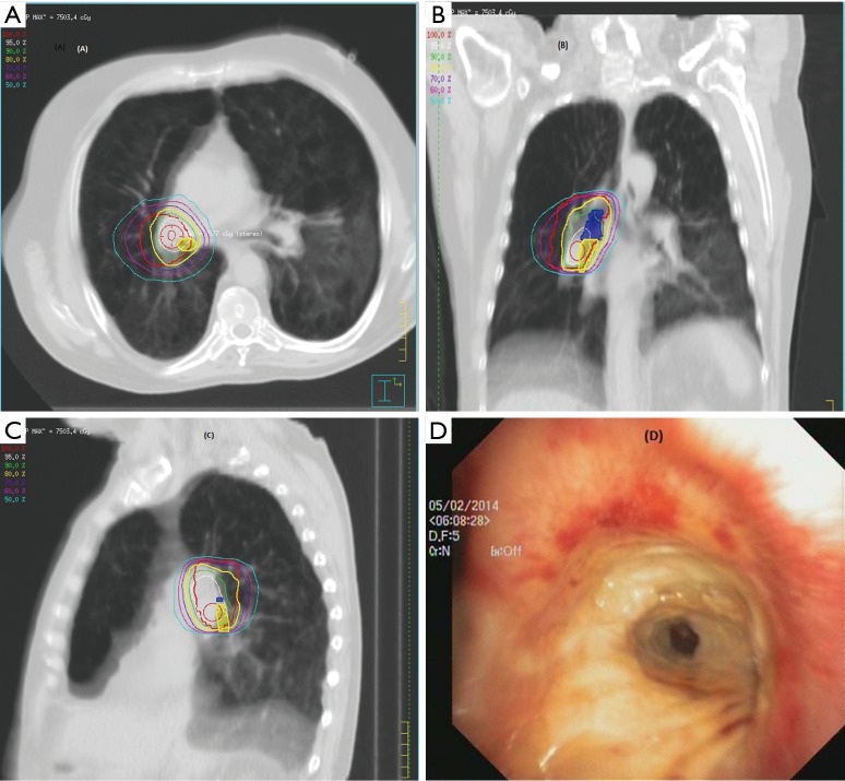
Figure 2.
Transversal, coronal and sagittal slices of the treatment planning computer tomography and corresponding endoscopic finding in the patient who developed bronchial necrosis after SABR for the central primary lung lesions (PLLs) in right hilum. Transversal (A) coronal (B) and sagittal (C) slices showed that a significant portion of the right main bronchus (blue) and the right intermediate bronchus (yellow) were within the planning target volume (PTV) and received dose maximum of more than 70 Gy. In the distal right main bronchus (D), the bronchoscopy revealed an area of hypervascularization above a circular area of necrosis that was located in the main right intermediate bronchus and lemon-yellow discolored necrotic cartilage.

