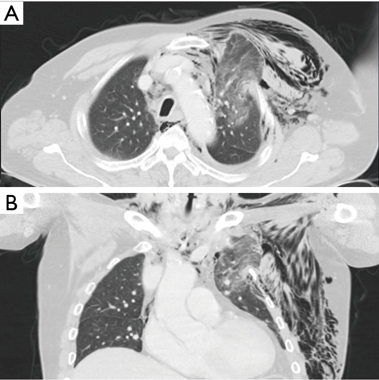
Figure 3.

Computed tomography performed three days after trauma shows herniation of the left upper lung with pneumomediastinum and a small left pneumothorax. Soft tissue emphysema was also evident at the left chest wall. A, axial view; B, coronal view.
