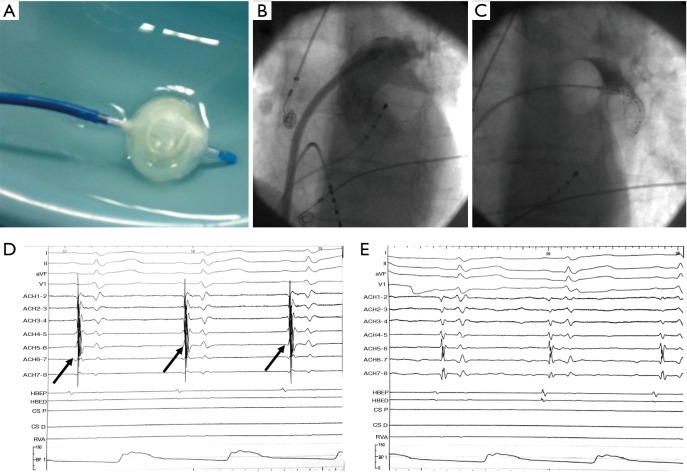
Figure 2.
A case of left upper pulmonary vein (PV) isolation is illustrated with use of the second generation cryoballoon (A). The anatomy of the PV is delineated via contrast injection (B) and then the vein is occluded with the balloon positioned at the entrance (see stasis of the contrast injected distally into the vein via the tip of the balloon in C). After completion of 3-minute freezing, the PV potentials (arrows, D) recorded prior to cryoenergy application are eliminated thus confirming successful isolation of this PV (E).

