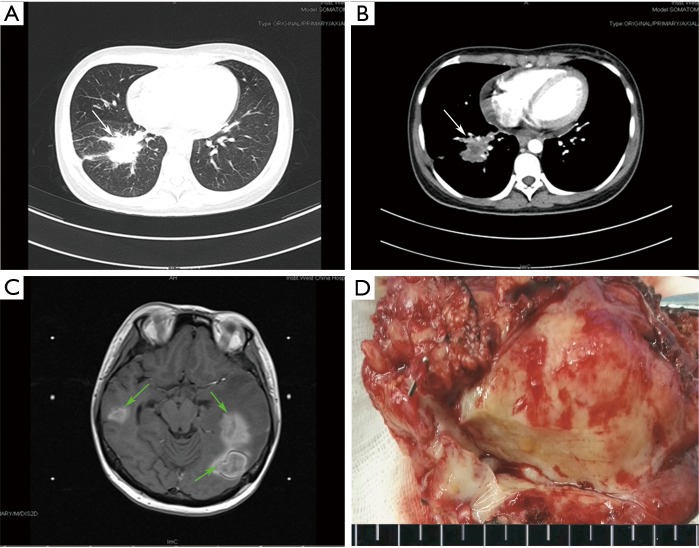
Figure 1.
Computed tomography (CT) and macroscopic imaging data of the patient with intracranial metastasis (Case 2) (A,B). CT examinations of the patient before the resection. A solid tumor (Marked by the white arrows) in the right lower lobe of the lung with deep lobulated sign, pleural indentation and markedly inhomogeneous enhancement showed on CT imaging; (C) 5 months after surgery, magnetic resonance imaging (MRI) showed a few of subcortical nodules with obvious peripheral edema (Marked by the green arrows) which were considered as intracranial metastasis; (D) a macroscopic image of the lobectomy specimen showed that the tumor was circumscribed, and whitish-yellow in color on the cut-surface.

