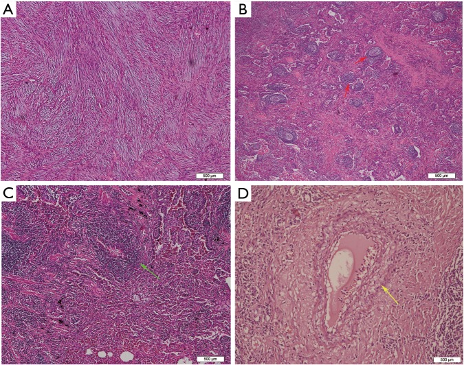
Figure 2.
Hematoxylin and eosin staining in IMT and IgG4-related IPT. (A) Original alveolar structures were totally destroyed by intersecting fascicles of spindle tumor cells in IMTs; (B) tumor cells were admixed with numerous lymphocytes, plasma cells and lymphoid follicles (red arrows) in IgG4-related IPTs; (C) a large number of inflammatory cells were infiltrative and involves the small bronchi (green arrow) in IPTs; (D) inflammatory infiltration of vessel walls or obstructive phlebitis (yellow arrow) were only observed in the tumors diagnosed as IgG4-related IPT.

