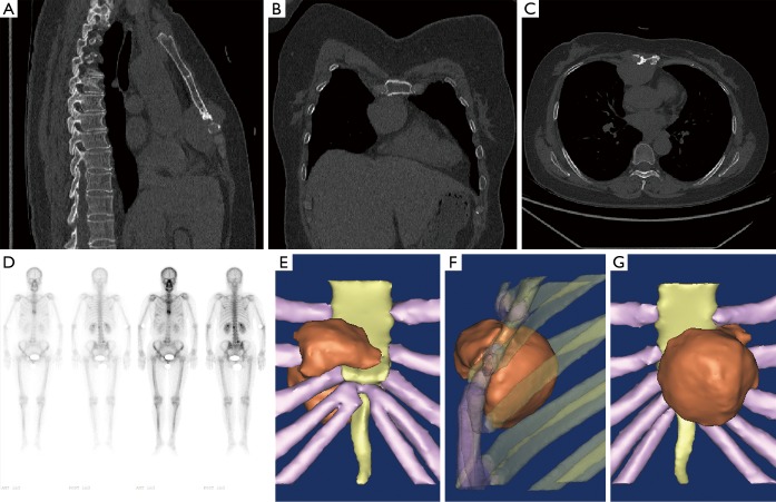
Figure 1.
Lesion on the CT, bone scintigraphy and 3D reconstruction: (A,B,C) a soft tissue mass (approximately 3.7 cm × 4.7 cm × 5.8 cm) located in the anterior mediastinum and anterior thoracic wall. The mass was likely to be malignant thymoma. The adjacent sternum was destroyed by the tumor; (D) bone scintigraphy revealed a nuclide focus on the distal mesosternum, first lumbar and fifth lumbar. Furthermore, these abnormal locations were suspected to be bone metastases; (E,F,G) 3D reconstruction displayed the certain appearance and location of the tumor. An overall demonstration was recorded in the short video (Figure 2).

