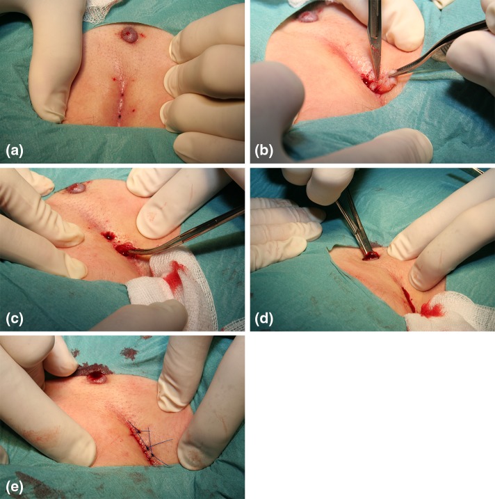
Fig. 1.
a Pilonidal sinuses and a proximal lateral tract. The area is infiltrated with local anaesthetic. b Pilonidal sinuses in the midline are excised through separate incisions with minimal margin. c All hairs and possible granulation tissue is removed with hemostat or a surgical spoon. d The tract is also searched and cleansed from hairs, using a hemostat. e In contrast to the original method the wound is closed primarily. The lateral tract is left open for drainage

