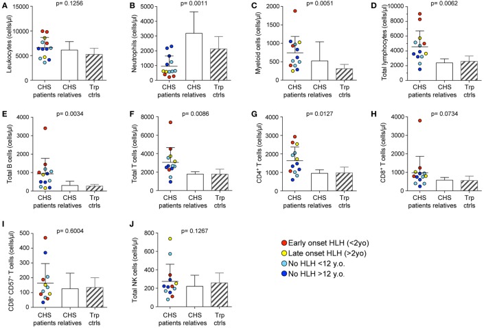
Figure 1.
Peripheral blood cell numbers in Chediak–Higashi syndrome (CHS) patients. (A) Total leukocyte, (B) neutrophil, (C) monocyte, (D) lymphocyte, (E) CD19+ B cell, (F) CD3+ T cell, (G) CD3+CD4+ T cell, (H) CD3+CD8+ T cell, (I) cytotoxic CD3+CD8+CD57+ T cell, and (J) bulk CD3−CD56+ NK cell numbers were enumerated in peripheral blood from 13 CHS patients. The patients are color-coded according to whether they presented with early-onset hemophagocytic lymphohistiocytosis (HLH) (<2 years), late-onset HLH (>2 years), or no HLH diagnosis, as indicated. Patient cell numbers were compared to those of healthy relatives and transport controls, as indicated. Columns depict mean values, bars indicate SD. Non-parametric one-way ANOVA Kruskal–Wallis tests are reported as exact p values.

