Figure 2.
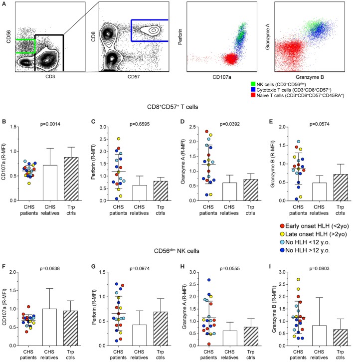
Expression of granule constituent proteins in cytotoxic lymphocyte subsets of Chediak–Higashi syndrome (CHS) patients. Gating strategy and representative dot plots of various lysosomal markers are shown in (A). Peripheral blood mononuclear cells were labeled for intracellular granule constituent proteins as well as surface proteins to distinguish [(B–E); n = 18 patients] CD3+CD8+CD57+ cytotoxic T cell and [(F–I); n = 20 patients] CD3−CD56dim NK cells, as indicated. Plots depict relative median fluorescent intensities of (B,F) CD107a, (C,G) perforin, (D,H) granzyme A, and (E,I) granzyme B in CHS patients, healthy relatives and transport controls, as indicated. The patients are color-coded according to whether they presented with early-onset HLH (<2 years), late-onset HLH (>2 years), or no HLH diagnosis, as indicated. Columns depict mean values, bars indicate SD. Non-parametric one-way ANOVA Kruskal–Wallis tests are reported as exact p values.

