FIG 1.
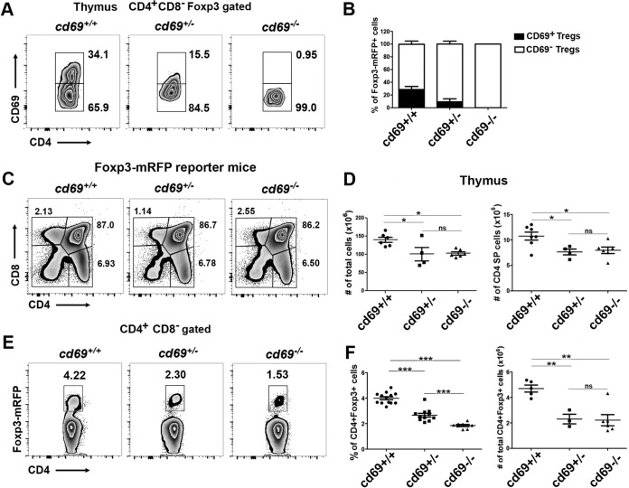
CD69 expression is required for thymus-derived Treg cell homeostasis in adult mice. (A) Density plots showing CD69 expression in CD4+ CD8− Foxp3+-gated thymocytes from 8- to 12-week-old Foxp3-mRFP/cd69+/+ (wild-type), Foxp3-mRFP/cd69+/− (heterozygous), and Foxp3-mRFP/cd69−/− (deficient) reporter littermates. Numbers indicate the proportions (percentages) of gated cells. (B) Bar chart showing the percentages (±standard deviations) of CD69+ and CD69− tTreg cells within the thymus of the indicated reporter mice. (C) Flow cytometry analysis of thymocyte subsets in 8- to 10-week-old reporter littermates. The percentages of thymus-derived T cell subsets are shown. (D) Cellularity of the thymus (left) and total numbers of CD4SP cells (right) in reporter littermates. (E) Analysis of endogenous Foxp3 expression in tTreg cells in the thymuses of reporter littermates. (F) Percentages (left) and total cell numbers (right) of gated CD4+ CD8− Foxp3+ tTreg cells in adult reporter littermates. Data are from at least 7 litters with 3 to 12 littermates each. Totals of 16 Foxp3-mRFP/cd69+/+ (wild-type), 11 Foxp3-mRFP/cd69+/− (heterozygous), and 12 Foxp3-mRFP/cd69−/− (deficient) mice were analyzed. Error bars show standard deviations. Data were evaluated by ANOVA followed by Bonferroni's multiple-comparison test. *, P < 0.05; **, P < 0.01; ***, P < 0.001; ns, not significant.

