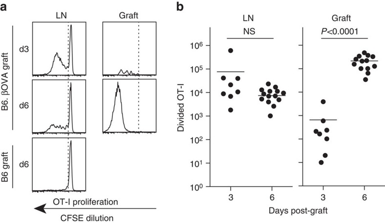
Figure 1. CD8+ T cells are primed in draining LN then expand at the site of inflammation.
Divided OT-I cells (viable CD45.1+CD8+Vα2+ gate) in draining renal LN and graft 3 or 6 days after receipt of a single graft of 400 B6.βOVA islets. (a) Representative flow cytometry plots. Position of the undivided OT-I peak was determined using ‘no antigen' control of a B6 islet graft. (b) Total number of divided OT-I in renal LN and graft where each point represents an individual mouse. Pooled data from seven independent experiments: n=8 graft recipients at day 3 and n=14 graft recipients at day 6. One day 6 graft was lost due to a flow cytometer malfunction. Horizontal bars are means, P values were calculated by unpaired, two-tailed t-test with Welch's correction.

