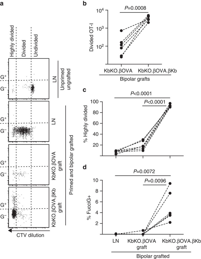
Figure 6. Cognate interaction with islet parenchymal β cells drives CD8+ T-cell proliferation.
FucciOT-I response to grafts in KbKO BM into B6 host mice in which host hematopoietic cells lack H-2Kb expression. Grafted mice received peptide-coated spleen cells on the day of grafting in order to initiate OT-I priming. (a) Representative flow cytometry plots (gated on viable CD45.1+CD8+Vα2+ lymphocytes). Upper panel shows lack of division and FucciG expression in quiescent OT-I in LN of a mouse that was neither grafted nor primed. Lower three panels show reponses in a bipolar grafted and primed mouse: draining renal LN, KbKO.βOVA and KbKO.βOVA.βKb grafts. Divided cells in grafted mice were divided into two sectors with the highly divided cells falling into the sector in which CTV was diluted beyond the limit of detection. (b) Total divided FucciOT-I in KbKO.βOVA and KbKO.βOVA.βKb bipolar grafts, P values calculated by two-tailed ratio paired t-test. (c) %highly divided and (d) % FucciG+ OT-I in draining renal LN and grafts of bipolar grafted mice. P values were calculated by two-tailed paired t-test. Results for individual mice are connected by dashed lines, n=6 recipient mice pooled from two independent experiments.

