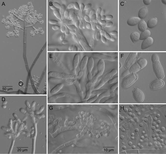
FIG 8.
Microscopic features of B. fragariae. (A) Large and small macroconidiophores in direct comparison. (B) Details of conidiogenesis on large macroconidiophores. (C) Conidia produced from large macroconidiophores. (D and E) Small macroconidiophores and details of conidiogenesis. (F) Conidia produced from small macroconidiophores. (G) Microconidiophore. (H) Microconidia. Panels B, C, E, F, G, and H are at the same scale.

