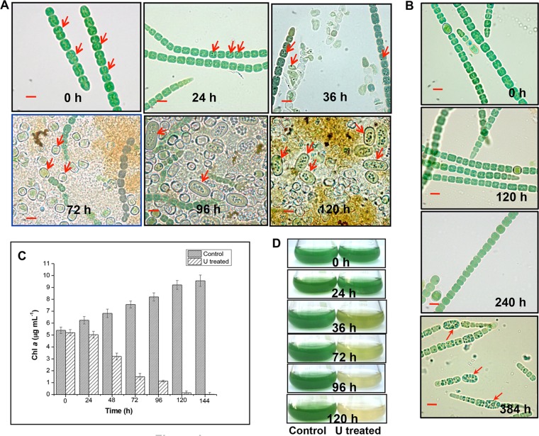
FIG 1.
Cell lysis, chlorosis, and akinete differentiation in U-exposed A. torulosa culture. Mid-log-phase cells at the equivalent of 0.2 mg (dry weight) ml−1 were exposed to 100 μM U (A) or unexposed to U (B) at pH 7.8 and were observed under a microscope under bright-field illumination (magnification, ×1,500; bars indicate 5 μm) at regular intervals. The data are a representative of three biological replicates. The red arrows in (A) show vegetative cells at 0 h, the dense dark granules formed as a result of colocalization of uranium with polyphosphate bodies at 24 h and 36 h in vegetative cells, heterocysts at 72 h, and akinetes at 96 h and 120 h. Red arrows in (B) indicate the differentiating akinetes at 384 h of incubation under control, uranium-unexposed phosphate-limited conditions. (C) Growth of U-unexposed control cells or cells exposed to 100 μM uranyl carbonate is represented by chlorophyll a (Chl a) contents. (D) The flasks containing control or U-challenged A. torulosa cultures corresponding to those mentioned in panel C.

