FIG 4.
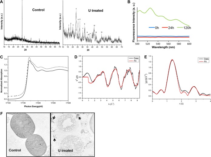
Characterization of bioprecipitated uranium. (A) XRD spectra of A. torulosa cells before and after 120 h of exposure to 100 μM uranyl carbonate. (B) Time-resolved fluorescence spectra of A. torulosa cells following 0 h, 24 h, and 120 h of uranium confrontation. The peaks at 505, 526, 550, and 575 nm, characteristic of chernikovite/meta-autunite, were observed for 120-h uranyl-exposed cells. (C) Normalized uranium LIII edge XANES spectra of 10−3 M U(VI) in 1 M HClO4 (dotted line) and 120-h uranyl-exposed cells (solid line). (D and E) Uranium LIII edge k2-weighted EXAFS spectrum (D) and corresponding FT (E) of uranium complexes formed by 120-h U-exposed A. torulosa cells. (F) Transmission electron micrographs of thin sections of A. torulosa cells before and after 120 h U exposure. The uranyl phosphate precipitates (indicated with arrows) were found to be scattered around the degraded cell aggregates. Uranyl composition of the precipitates was previously confirmed by energy dispersive X-ray fluorescence (EDXRF) spectroscopy, which revealed all components of uranium L (UL) X rays, i.e., ULl, ULα, ULβ1, and ULβ2.

