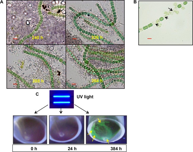
FIG 6.
Features of regenerated A. torulosa cells and stability of the biomineralized uranyl following cell regeneration. (A) Regenerated cells stained with toluidine blue were observed using bright-field microscopy in a Carl Zeiss Axioskop 40 microscope (Carl Zeiss, Germany) (magnification, ×1,500; bars indicate 5 μm) following 240 to 384 h of uranium exposure. No polyphosphates were visualized in these regenerating cells. (B) Regenerated cells exposed to 100 μM U for 5 min under shaking conditions were observed using bright-field microscopy (bar indicates 5 μm). The regenerated cells showed lysis (marked by black arrows) suggestive of their sensitivity to U toxicity. (C) Cells of A. torulosa were incubated with 100 μM U for 0 h, 24 h, or 384 h and the respective cell pellets were exposed to UV light and photographed. The uranyl precipitates at the bottom of the regenerated biomass at 384 h displayed a distinct green fluorescence (marked by yellow arrows) consistent with autunite mineral.

