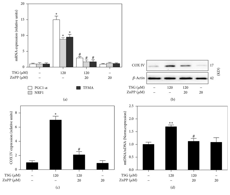
Figure 5.
The mitochondrial biogenesis induced by TSG was regulated by activation of HO-1. RAW264.7 cells were treated with TSG (120 μM) for 6 hours in the presence and absence of ZnPP (20 μM). (a) After treatment, mRNA expressions of PGC-1, NRF1, and TFAM were detected by Real-Time PCR. (b) Western blot analysis was used to assess the expression of mitochondrial complex IV in whole cell lysates. (c) The relative COX-IV protein level was analyzed by densitometry. (d) Quantitative PCR was performed to evaluate the mtDNA content with beta2-microglobulin and cytochrome b served as probes specific for nuclear DNA and mtDNA. Relative ratios of mtDNA and nDNA contents were analyzed. All measurements were expressed as the mean ± SEM of triplicate independent experiments (n = 3). ∗P < 0.05 and ∗∗P < 0.01 versus control group; #P < 0.05 versus TSG treatment group.

