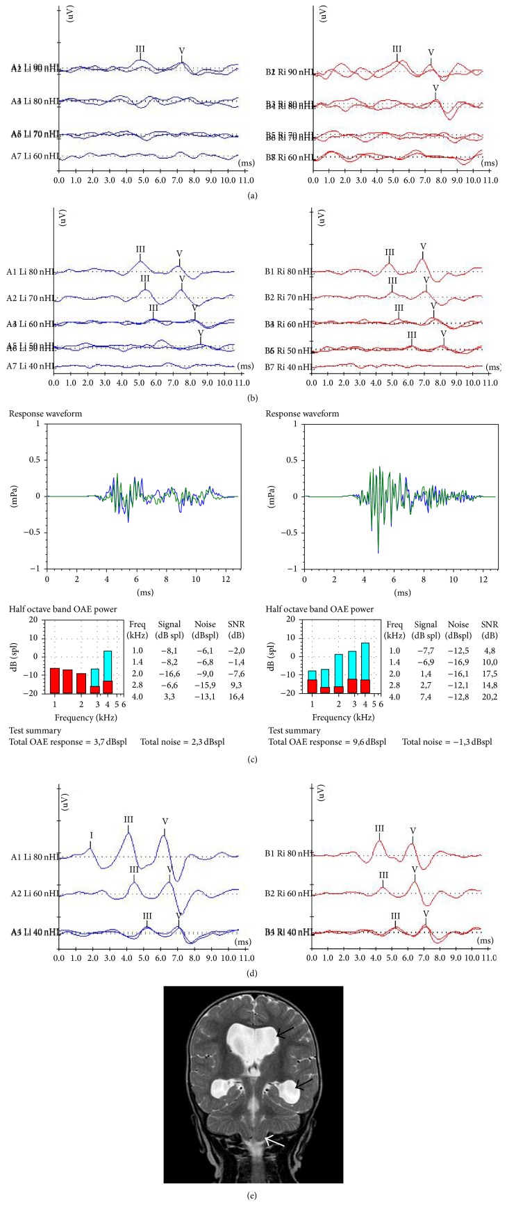
Figure 3.
Serial ABR measurements, otoacoustic emissions and MRI findings of an infant with type I Chiari malformation. (a) ABR recordings at the initial hearing assessment (age of 2.5 months). Waveforms were obtained at 90 dBnHL on the left and 80 dBnHL on the right ear. (b) ABR findings 5 months later (age of 7.5 months). Waveforms were elicited bilaterally at 50 dBnHL. (c) At the same time, normal otoacoustic emissions were recorded on the right side and partial response on the left. (d) Last ABR session after 10 months (age of 18 months). Typical ABR waveforms were recorded at 40 dBnHL bilaterally, a finding which corresponds to “normal” ABR threshold and which is considered a strong indication of normal hearing. (e) Coronal MRI image of the same infant at age of 7 months, depicting enlargement of lateral ventricles (black arrows) and herniated cerebellar tonsils (white arrow). In this case, the ABR thresholds recovered completely.

