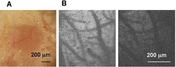
Fig. 1.

Thinned-skull cranial window (TSCW) and cortical light exposure. (A) Low-magnification bright field image of TSCW. (B) NADH autofluorescence through TSCW. Two photon microscopy images (Ex 740 nm/Em 450 m) before (left) and after (right) light exposure through a 20× microscope objective (90 W halogen bulb, 2.5 min).
