
Fig. 10.
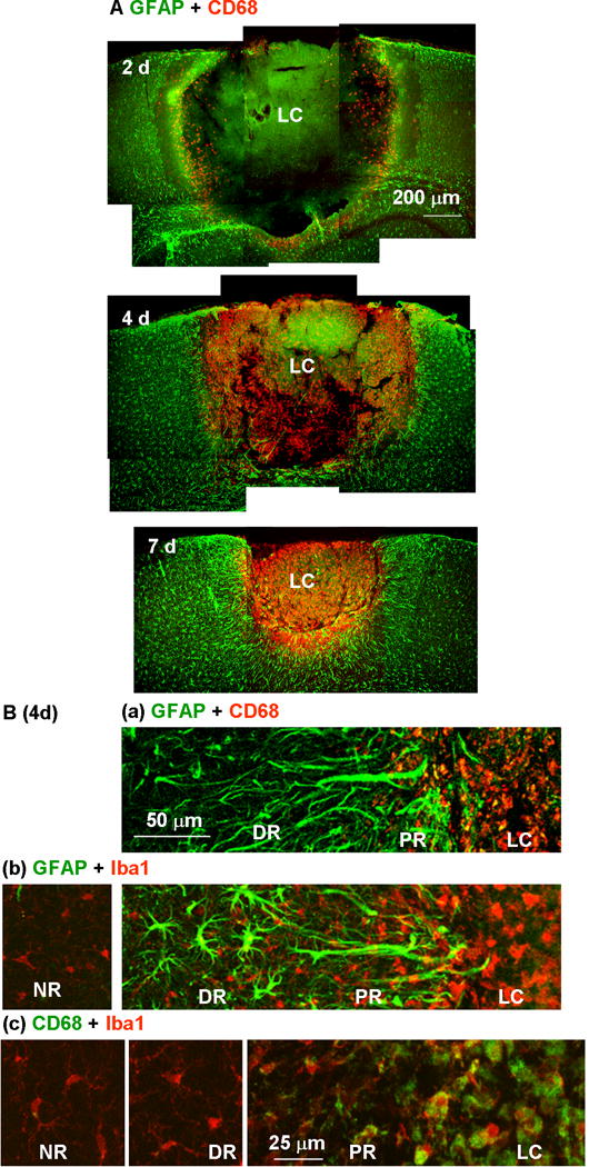
Restriction of microglial distribution by the proximal reactive astrocyte cell layer. (A) Reactive astrocytes and activated microglia in the injured cortex were stained with anti-GFAP (green) and anti-CD68 (red) antibodies, respectively, 2, 4, and 7 days after photo injury. (B) Higher magnification images on day 4 illustrated the distribution of microglia at different activation states in the lesion core (LC), proximal region (PR), distal region (DR), and normal region (NR). Microglia were stained with anti-CD68 (a) or the pan-microglial marker, Iba1 (b). The label colors are indicated above each set of panels. In the panels in (c), microglia stained for CD68 (green) and Iba1 (red) were further magnified for comparison of structures in different regions.