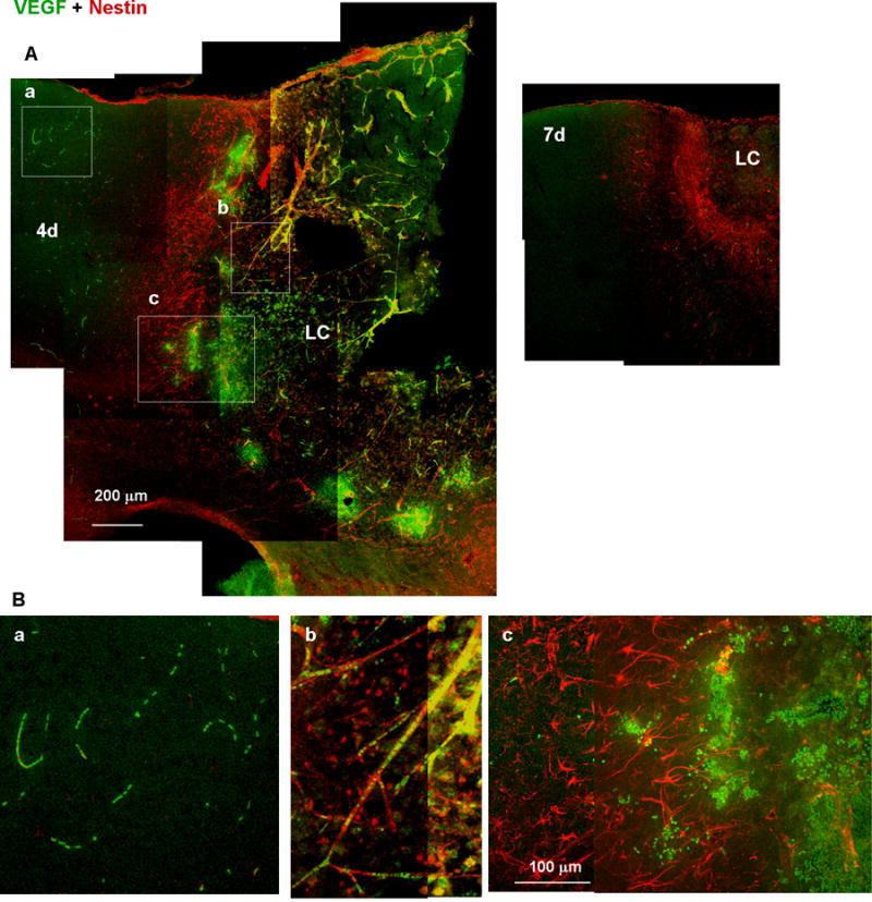
Fig. 11.

Accumulation of VEGF-expressing leukocytes at the early stages of wound healing. (A) Low-magnification images of tissue labeled with anti-VEGF (green) and anti-nestin (red) antibodies. VEGF immunoreactivity was distributed in both the distal region (a), which is outside of the nestin-positive proximal reactive astrocyte layer, and in the lesion core (LC; b and c), which is inside of the reactive astrocyte layer, 4 days after injury (4d), but undetectable on day 7 (7d). (B) High-magnification images of the boxes in (A). VEGF immunoreactivity was localized in leukocytes in the presumed distal region capillaries (a) or LC vessels (b), or accumulated at the border between the reactive astrocyte layer and the LC.
