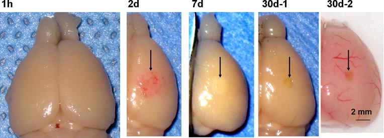
Fig. 2.

Low-magnification photographs of lesioned brains after photo injury 1 h – 30 days after injury. As mentioned in Materials and Methods, photo injury was induced in the right somatosensory cortex. Brains were dissected after paraformaldehyde fixation by transcardiac perfusion (1 h, 2 d, 7 d, and 30d-1) or without fixation (30d-2). Post-injury tissue changes are indicated by arrows in subsequent frames.
