Fig. 3.

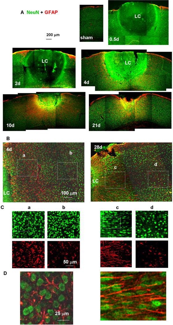
Tissue sections showing the lesion core (LC) and perilesional reactive astrocytes. Neuronal nuclei and astrocytes were stained with anti-NeuN (green) and anti-GFAP (red) antibodies, respectively. (A) Low-magnification images of a sham-treated (no light injury, fixed 4 days after skull thinning) mouse cortex underlying TSCW and injured cortex at 0.5, 2, 4, 10, and 21 days after photo injury. (B) Higher magnification images of the perilesional region on days 4 and 28. The NeuN (green)- and GFAP (red)-positive structures in Boxes (a) – (d) are further magnified in (C). On day 4, the morphology of neuronal nuclei was similar between the regions (a) proximal and (b) distant to LC. On day 28, neuronal nuclei showed pronounced elongation running along the direction of GFAP fibers in (c) the proximal region, but there were no significant differences from day 4 in (d) the distal region. In (D), the boxed regions shown in (C) are further magnified and merged to emphasize the elongation of NeuN staining nuclei along the GFAP-staining fibers. In this figure only, GFAP labeling is shown in red, reserving green for NeuN, which gave a clearer definition of nuclear shape.
