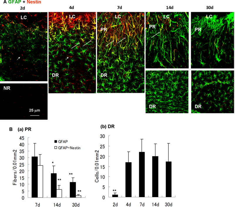
Fig. 7.

Time-dependent changes of reactive astrocytes after photo injury. (A) Time course of nestin (red) and GFAP (green) immunoreactivity in reactive astrocytes (days 2, 4, 7, 14, and 30). Perilesional GFAP expression was upregulated more than the normal region (NR) by day 2. Structural difference of reactive astrocytes between the lesion core (LC), the proximal region (PR) and the distal region (DR) was established by day 7. Nestin-expressing reactive astrocytes were clearly identified by day 4, reached the maximum level on day 7, and decreased to below the limit of detection by day 30. On day 4, the morphology of reactive astrocytes was uniform, whereas nestin-expressing reactive astrocytes indicated by arrows were localized in the proximity of the LC. On days 14 and 30, the DR is shown in separate pictures because the region of high-density reactive astrocytes was remote from the LC, reflecting the enlargement of the PR. Arrowheads in day 2 and 4 indicate nestin-positive and capillary-like structures without GFAP immunoreactivity, presumably reflecting proliferating endothelial cells in damaged vessels. (B) Cell densities of reactive astrocytes. Density of radial fibers in PR (GFAP-positive or GFAP/nestin-positive) or cell bodies of GFAP-positive reactive astrocytes in DR were counted in areas of 100 μm × 100 μm. Means ± SD (n = 3 images at each time point), ** P < 0.01, * P < 0.05 ANOVA and Dunnett’s test for multiple comparisons with 7 days.
