Fig. 8.

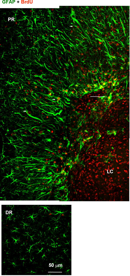
Proliferative nature of proximal reactive astrocytes. Tissue was labeled with anti-GFAP (green) and anti-BrdU (red) antibodies. BrdU was administrated between 1 and 6 days after photo injury and animals were sacrificed on day 7. Fluorescence images of the lesion core (LC) and proximal region (PR) (upper panels), and distal region (DR) (lower). The BrdU-positive but GFAP-negative nuclei in the LC likely correspond to microglia (see also Fig. 10). In the PR, the majority of GFAP-positive structures were co-localized with BrdU-positive nuclei. Arrowheads indicate BrdU-positive reactive astrocytes extending processes radially. In the DR, no significant co-localization of BrdU and GFAP was found. The arrow indicates the extension of GFAP-positive structure into LC.
