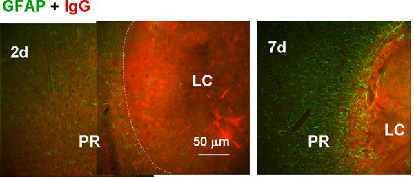
Fig. 9.

Blockade of blood component diffusion by the proximal reactive astrocyte layer. Reactive astrocytes and blood components were stained with anti-GFAP and anti-IgG antibodies, respectively. On day 2, IgG immunoreactivity was diffuse outside of the LC (approximate boundary indicated by the dashed line); whereas on day 7, it was limited to within the LC, which was surrounded by reactive astrocytes.
