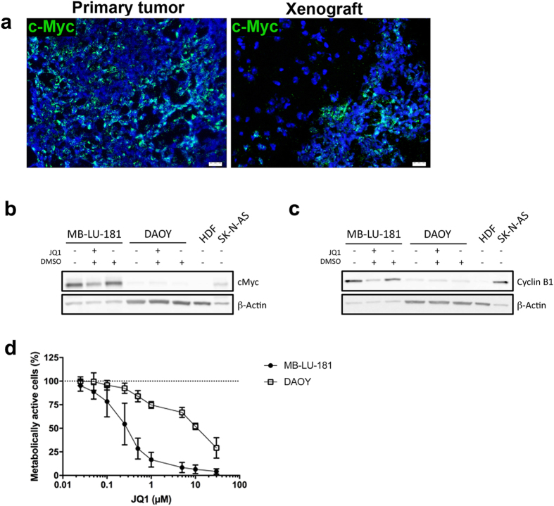
Figure 3. Effect of MYC inhibition in patient-derived Group 3 medulloblastoma.
(a) MYC expression is demonstrated in primary tumour and xenografts with in situ hybridization. Scale bar is 20 μm. Western blotting detecting c-Myc (b) and cyclin B1 (c) in protein extracts isolated from MB-LU-181 cells JQ1 treatment. SK-N-AS neuroblastoma cells and human dermal fibroblasts (HDF) cells were included as positive and negative control for c-Myc expression, respectively. β-actin was used to ensure equal sample loading. (d) Cell viability measurement using WST-1 of (d) MB-LU-181 and DAOY cells treated with the indicated concentrations of JQ1 for 72-hours. IC50 values between MB-LU-181 and DAOY cells were significantly different (p < 0.0001 t-test; 0.27 μM in MB-LU-181 versus 10 μM in DAOY). Mean with SD from three independent experiments are displayed.

