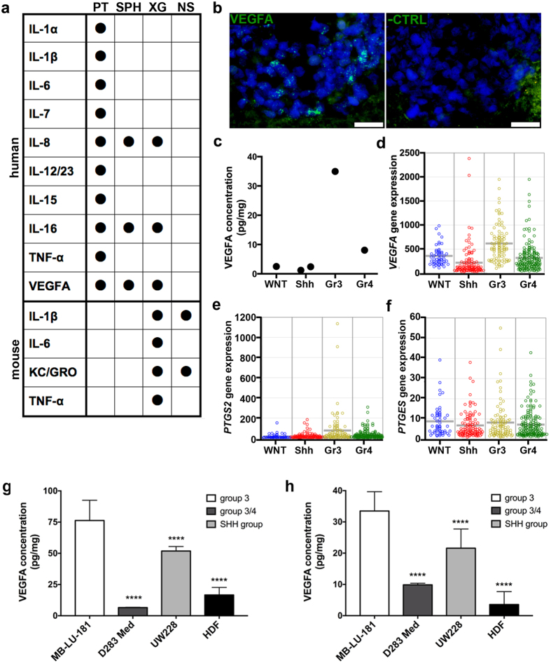
Figure 5. Inflammatory factors in primary tumour, patient-derived sphere cultures and xenografts.
(a) A panel of inflammatory factors was analysed with MesoScale arrays. Values above LLOQ are indicated with a black dot. PT, primary tumour; XG, xenograft; SPH, cultured spheres; NS, NOD-scid cerebellum. (b) VEGFA is expressed in xenografted medulloblastoma cells, demonstrated by in situ hybridization. To the left, VEGFA probe; to the right, negative control probe on parallel section. Scale bar is 20 μm. (c) Mesoscale analysis of VEGFA protein levels in 5 primary medulloblastoma tissues. Median of duplicate values is shown. Gene expression levels of (d) VEGFA, (e) PTGS2 (COX-2) and (f) PTGES (mPGES-1) in medulloblastoma patients (WNT, n = 53; Shh, n = 112; Group 3, n = 94; Group 4; n = 164). VEGFA expression in (g) supernatants and (h) cell extracts from MB-LU-181, D283 Med (group 3/4), UW228 (SHH group) and human dermal fibroblast (HDF) cells were analysed with VEGFA-ELISA. Significant differences compared to MB-LU-181 are marked by asterisks (p < 0.0001 (****); one-way ANOVA with Bonferroni as post-test).

