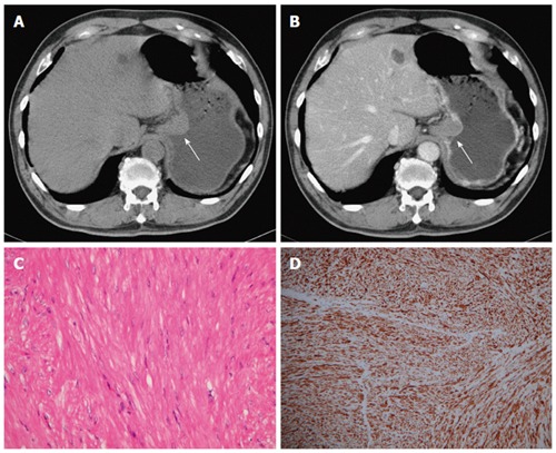
Figure 3.

Leiomyoma. A 70-year-old man had no symptoms. A and B: Pre- and post-contrast-enhanced axial computed tomography scans showing the leiomyoma at the gastric cardia (arrow), with an intraluminal growth pattern and homogeneous, poor enhancement. Note the intact enhancing mucosa, indicating the submucosal lesion; C: High-power photomicrograph (original magnification, × 200; HE stain) showing paucicellular spindle cells with low or moderate cellularity, arranged in perpendicularly oriented fascicles; The tumor cells were positive for smooth muscle actin (D) and negative for CD34 and CD117 (not shown).
