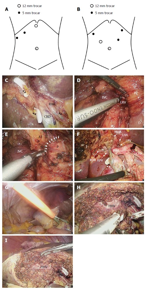
Figure 3.

Surgical procedure for laparoscopic gallbladder bed resection. A: Position of trocars in laparoscopic gallbladder bed resection (LCGB) with D1 lymphadenectomy; B: Position of trocars in LCGB with D2 lymphadenectomy; C: The cystic artery and duct are cut at their origin; D: Kocher’s mobilization; E: Lymph node dissection around the posterosuperior region of the pancreas head. Arrow indicates the boundary between the pancreatic parenchyma and surrounding adipose tissues; F: Completion of D2 lymphadenectomy; G: Performance of the Pringle maneuver with an extracorporeal tourniquet; H: Transection of the liver parenchyma by the clamp crushing method; I: After the gallbladder bed resection. RHA: Right hepatic artery; CBD: Common bile duct; Arrowhead: Stump of the cystic duct; Dotted arrow: Stump of the cystic artery; P: Pancreatic head; D: Duodenum; IVC: Inferior vena cava; LRV: Left renal vein; AT: Adipose tissues; GDA: Gastroduodenal artery; CHA: Common hepatic artery; PHA: Proper hepatic artery; LHA: Left hepatic artery; MHA: Middle hepatic artery; PV: Portal vein; LPV: Left portal vein; RPV: Right portal vein; MHV: Middle hepatic vein.
