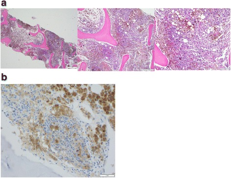
Fig. 1.

Image of patient’s bone marrow biopsy, confirming metastatic melanoma to bone with corresponding PD-L1 staining. Panel a shows images of the patient’s bone marrow with extensive involvement by metastatic melanoma. Panel b shows corresponding bone marrow sample with tumor cells staining positive for PD-L1 (Vendor: Cell signal, Clone: E1L3N, Dilution: 1:500)
